
ANATOMY OF THE EYE
| |||||||||||||||
|
|
| |||||||||||||


Understanding how the eye works and what role the cornea plays in this process will help to understand keratoconus.
The eye captures and focuses light like a camera. Here is a step-by-step explanation of how the eye works to provide you with vision:
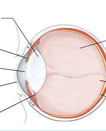
- Light enters the eye through the cornea (the clear, dome-shaped surface that covers the front of the eye).
- From the cornea, the light passes through the pupil. The amount of light passing through is regulated by the iris, or the colored part of your eye.
- From there, the light then hits the lens, the transparent structure inside the eye, which focuses light rays onto the retina.
- Finally, it reaches the retina, the light-sensitive nerve layer that lines the back of the eye, where the image appears inverted.
- The optic nerve carries signals of light, dark, and colors to the area of the brain (the visual cortex), which assembles the signals into images (our vision).
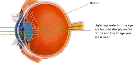
With normal vision, the cornea and the lens focus light directly onto the retina. Light rays entering the eye are focused sharply on the retina and the image you see is clear.
Because the keratoconus cornea is irregular and cone shaped, light rays enter the eye at different angles, and do not focus on one point on the retina, but on many different points causing a blurred, distorted image.